A chemo port, also known as a port-a-cath or an implanted port, is a small medical device placed under the skin that allows for easy access to the bloodstream during chemotherapy treatment.
It consists of two main components: a catheter, which is a thin, flexible tube, and a port, which is a reservoir that connects to the catheter.
The catheter is inserted into a large vein, typically in the neck and the port is implanted just beneath the skin, usually in the chest area.
The procedure often requires general anesthesia.
Once the port is in place, it eliminates the need for repeated needle sticks, and is recommended when chemotherapy or targeted therapy is advised, chemotherapy drugs can be directly administered through the port.
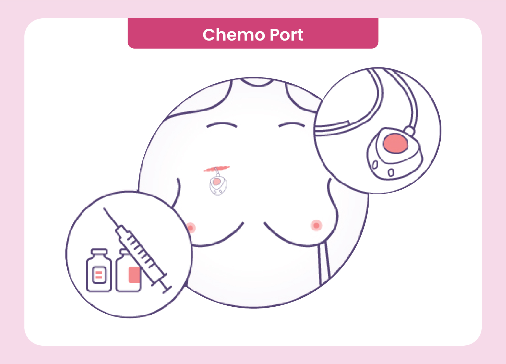
The port is typically accessed using a special needle, called a Huber needle, which is inserted through the skin and into the port.
The port is designed to be long-lasting and can remain in place for the duration of the chemotherapy treatment, which can last several months.
It is important to keep the chemo port clean and dry to prevent infections, and regular flushing and maintenance are necessary to ensure its proper functioning.
Removal of the chemo port is a relatively simple procedure and is usually done under local anesthesia. once the chemotherapy treatment is completed.
Sentinel lymph node biopsy or axillary sampling are two techniques used in the staging and defining if the lymph nodes under the armpit or axilla are positive. This is required for and treatment planning for certain types of cancer, particularly breast cancer.
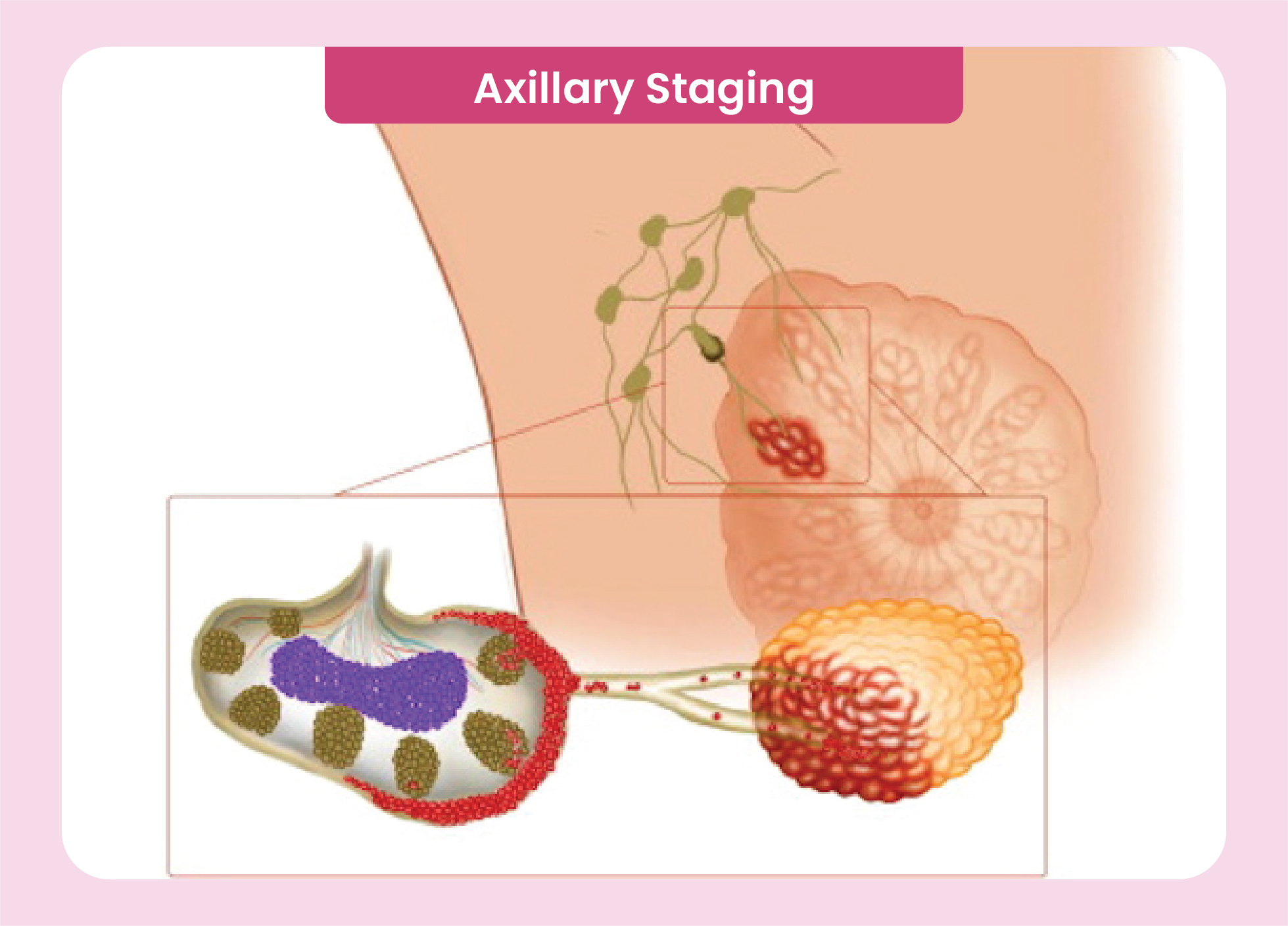
Axilla mapping is performed to determine if cancer cells have spread to the lymph nodes in the axillary (underarm) region from the primary tumor site, usually the breast.
The lymph nodes in the axilla are the first ones to which cancer cells are likely to spread from the breast.
Sentinel lymph node biopsy :The procedure involves injecting a small amount of a dye (blue coloured, fluoresceine or radiocolloid) around the tumour.
The dye travels along the lymphatic vessels and accumulates in the first few lymph nodes, known as the sentinel lymph nodes, that receive drainage from the tumor.
The sentinel lymph nodes are then identified and surgically removed and sent to a pathologist for examination.
Pathological examination of the sentinel lymph nodes helps determine if cancer cells have spread to the axillary lymph nodes.
If cancer cells are found in the sentinel lymph nodes, additional treatment options such as axillary lymph node dissection is be considered.
Axilla mapping can by axillary sampling where, the surgeon removes 4-6 lymph nodes from the axilla based on certain anatomical structures and those are sent for pathological evaluation. The further procedure is same as described above.
These technique reduces the need for more extensive lymph node surgery, reducing potential complications and side effects associated with complete axillary lymph node dissection, like lymphedema (or swelling of the arm).
It is important to note that specific details of axilla mapping may vary depending on the individual case, the healthcare facility, and the specific protocol followed by the medical team.